Germ Cell Death
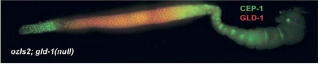
GLD-1 acts as a translational repressor of CEP-1/p53. Double staining with anti-CEP-1 antibodies and anti-GFP antibodies of germlines from gld-1(null); ozIs2 adult hermaphrodites expressing a GLD-1::GFP fusion protein.
As is true in all metazoa, C. elegans germ cells are both pluripotent and immortal, in that they give rise to all cell types in the subsequent generations, capable of forming entire organism. One conserved aspect of germline development is that in organisms as diverse as C. elegans and humans, a high percentage of developing oocytes die by apoptosis (programmed cell death) during or just after the pachytene stage of meiotic prophase. For instance, in the mammalian female ovaries, apoptosis occurs throughout fetal and postnatal life to claim up to 99.9 % of potential germ cells. Although most germ cell deaths take place prior to birth, apoptosis continues to decimate the number of oocytes through a process called follicular atresia during juvenile and adult life in mammals. Once the supply of oocytes has been exhausted through apoptosis and ovulation, the ovaries senesce, leading to infertility and, in humans, to menopause. How programmed cell death is selectively modulated so that some oocytes survive while others die remains a mystery.
In C. elegans somatic apoptosis is developmentally programmed by invariant cell lineage: always the same 131 of the 1090 somatic cells die by programmed cell death. In contrast, germline apoptosis does not follow a set pattern and appears to be an intrinsic part of the oogenesis program. Germ cells can activate apoptosis in response to developmental cues, but also in response to external cues, including genotoxic stress, bacterial infection and the failure of meiotic chromosomes to synapse. Recently it was shown that cisplatin, a chemotherapeutic drug, induces apoptosis in human germ cells which leads to ovarian failure and infertility. The high degree of similarity between worms and humans at the level of genes and biochemical pathways make C. elegans germ cells a particularly interesting tissue to study apoptosis and its regulation in response to developmental and environmental cues.
- Martin Keller
- Deni Subasic
- Cathy Zheng
Sendoel A, Maida S, Zheng X, Teo Y, Stergiou L, Rossi CA, Subasic D, Pinto SM, Kinchen JM, Shi M, Boettcher S, Meyer JN, Manz MG, Bano D, Hengartner MO. DEPDC1/LET-99 participates in an evolutionarily conserved pathway for anti-tubulin drug-induced apoptosis. Nat Cell Biol. 2014 Aug;16:812-20.
Kratz K, Schöpf B, Kaden S, Sendoel A, Eberhard R, Lademann C, Cannavó E, Sartori AA, Hengartner MO, Jiricny J:Deficiency of FANCD2-associated nuclease KIAA1018/FAN1 sensitizes cells to interstrand crosslinking agents. Cell 2010, 142: 77-88.
Sendoel A, Kohler I, Fellmann C, Lowe SW, Hengartner MO :HIF-1 antagonizes p53-mediated apoptosis through a secreted neuronal tyrosinase. Nature 2010, 465: 577-83.
Zermati Y, Mouhamad S, Stergiou L, Besse B, Galluzzi L, Boehrer S, Pauleau AL, Rosselli F, D'Amelio M, Amendola R, Castedo M, Hengartner M, Soria JC, Cecconi F, Kroemer G: Nonapoptotic role for Apaf-1 in the DNA damage checkpoint. Mol Cell 2007, 28:624-637.
Kritikou EA, Milstein S, Vidalain P, Lettre G, Bogan E, Doukoumetzidis K, Gray P, Chappell TG, Vidal M, Hengartner MO. C. elegans GLA-3 is a novel component of the MAP kinase MPK-1 signaling pathway required for germ cell survival. Genes Dev. 2006 Aug 15;20(16):2279-2292.
Schumacher B, Hanazawa M, Lee MH, Nayak S, Volkmann K, Hofmann R, Hengartner MO, Schedl T, Gartner A: Translational repression of C. elegans p53 by GLD-1 regulates DNA damage-induced apoptosis. Cell 2005, 120:357-368.